Bacterial spore defile is a critical technique in microbiology, crucial for identifying and examine bacterial spores. These spores are inactive, tough, and extremely resistant structures formed by certain bacteria under adverse conditions. They play a pivotal role in the survival and diffusion of bacterial species, making their designation crucial in various fields, including medicine, food safety, and environmental skill.
Understanding Bacterial Spores
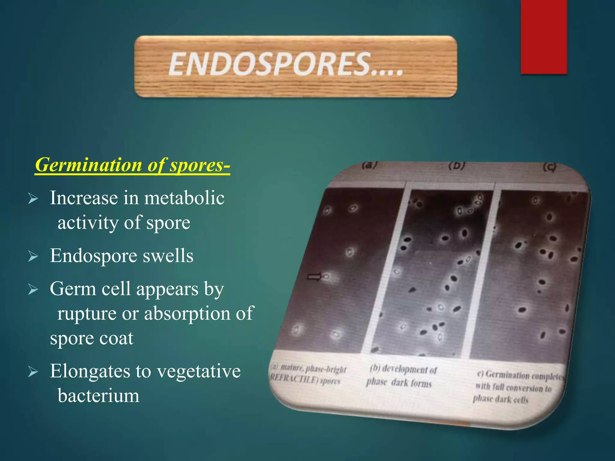
Bacterial spores are formed through a summons called sporulation, which occurs when bacteria skirmish unfavorable environmental conditions such as nutrient depletion, high temperatures, or the presence of toxins. Spores are characterize by their thick, protective coat and their power to remain inactive for extended periods, sometimes even centuries. When conditions amend, spores can germinate and revert to their vegetive state, resume normal bacterial activities.
Importance of Bacterial Spore Staining
Bacterial spore maculate is critical for several reasons:
- Identification of Spores: It helps in the designation of spore constitute bacteria, which is essential for symptomatic purposes in clinical and environmental settings.
- Research and Development: Understanding the formation and properties of spores is all-important for developing new antimicrobial agents and treatments.
- Food Safety: Many foodborne pathogens, such as Clostridium botulinum and Bacillus cereus, form spores that can foul food products, making spore defile important for ensuring food safety.
- Environmental Monitoring: Spores can persist in the environment for long periods, making spore staining a worthful instrument for monitor environmental contamination and assessing the effectiveness of decontamination procedures.
Types of Bacterial Spore Staining Techniques
Several defile techniques are used to visualize bacterial spores. The choice of technique depends on the specific requirements of the study and the characteristics of the spores being examined.
Schaeffer Fulton Method
The Schaeffer Fulton method is one of the most commonly used techniques for bacterial spore staining. This method involves a series of steps that tell spores from vegetive cells based on their defile properties.
The steps imply in the Schaeffer Fulton method are as follows:
- Prepare a smear of the bacterial acculturation on a microscope slide and grant it to air dry.
- Heat fix the smear by pass the slide through a flame various times.
- Stain the smear with malachite green for 5 10 minutes. This primary stain penetrates both the spores and vegetal cells.
- Rinse the slide with h2o to remove excess stain.
- Counterstain the smear with safranin for 1 2 minutes. Safranin stains the vegetative cells but does not penetrate the spores.
- Rinse the slide with water and blot dry.
- Examine the slide under a microscope. Spores will appear green, while vegetative cells will appear pink or red.
Note: The Schaeffer Fulton method is specially useful for place spores in fuse cultures, as it clearly differentiates spores from vegetive cells.
Dorner Method
The Dorner method is another technique used for bacterial spore staining. This method is similar to the Schaeffer Fulton method but uses different stains and a shorter staining time.
The steps involved in the Dorner method are as follows:
- Prepare a smear of the bacterial acculturation on a microscope slide and allow it to air dry.
- Heat fix the smear by passing the slide through a flame several times.
- Stain the smear with carbol fuchsin for 5 10 minutes. This primary stain penetrates both the spores and vegetative cells.
- Rinse the slide with water to remove excess stain.
- Counterstain the smear with methylene blue for 1 2 minutes. Methylene blue stains the vegetive cells but does not perforate the spores.
- Rinse the slide with water and blot dry.
- Examine the slide under a microscope. Spores will appear red, while vegetative cells will appear blue.
Note: The Dorner method is ofttimes preferred for its shorter staining time and the use of carbol fuchsin, which provides a more distinct color contrast between spores and vegetational cells.
Gram Staining
While not specifically designed for spore maculate, Gram tarnish can also be used to identify bacterial spores. This technique differentiates bacteria based on their cell wall characteristics, with spores frequently appearing as Gram positive structures.
The steps affect in Gram sully are as follows:
- Prepare a smear of the bacterial culture on a microscope slide and let it to air dry.
- Heat fix the smear by surpass the slide through a flame respective times.
- Stain the smear with crystal violet for 1 minute.
- Rinse the slide with water and apply iodine answer for 1 minute.
- Decolorize the smear with alcohol or acetone for a few seconds.
- Counterstain the smear with safranin for 1 2 minutes.
- Rinse the slide with h2o and blot dry.
- Examine the slide under a microscope. Spores will appear purple, while Gram negative cells will appear pink or red.
Note: Gram staining is a versatile technique that can be used for a extensive range of bacterial identification purposes, including the catching of spores.
Applications of Bacterial Spore Staining
Bacterial spore staining has legion applications in respective fields, including medicine, food safety, and environmental science.
Medical Diagnostics
In aesculapian diagnostics, bacterial spore maculate is used to identify spore forming pathogens that can make serious infections. for instance, Clostridium difficile is a spore forming bacterium that can induce severe gastrointestinal infections, particularly in hospitalized patients. Accurate designation of C. difficile spores is important for diagnosing and process infections.
Food Safety
In the food industry, bacterial spore tarnish is essential for ensuring the safety of food products. Many foodborne pathogens, such as Bacillus cereus and Clostridium botulinum, form spores that can survive harsh processing conditions and contaminate food. Spore tarnish helps in discover these pathogens and enforce allow control measures to prevent foodborne illnesses.
Environmental Monitoring
In environmental skill, bacterial spore staining is used to monitor the presence of spores in soil, h2o, and air. Spores can persist in the environment for long periods and can be indicators of environmental taint. Spore staining helps in assessing the effectivity of decontamination procedures and ensuring environmental safety.
Challenges and Limitations of Bacterial Spore Staining
While bacterial spore tarnish is a valuable technique, it also has respective challenges and limitations.
Staining Artifacts
One of the chief challenges in bacterial spore stain is the presence of tarnish artifacts, which can interfere with the accurate designation of spores. Artifacts can be caused by improper staining techniques, taint, or the front of other cellular structures that mimic spores.
Sensitivity and Specificity
Another limitation of bacterial spore tarnish is its sensibility and specificity. Some staining techniques may not be sensitive enough to detect low levels of spores, while others may produce false convinced results due to non specific tarnish. It is essential to opt the seize staining technique and optimize the stain conditions to insure accurate results.
Interpretation of Results
Interpreting the results of bacterial spore staining can be gainsay, specially in conflate cultures or complex samples. It requires a eminent stage of expertise and experience to accurately differentiate spores from other cellular structures and artifacts.
Future Directions in Bacterial Spore Staining
Advances in technology and enquiry are continually better bacterial spore stain techniques. Some of the hereafter directions in this field include:
Automated Staining Systems
Automated staining systems are being germinate to standardise and streamline the staining summons. These systems can reduce human fault, better consistency, and increase the throughput of spore maculate procedures.
Molecular Techniques
Molecular techniques, such as polymerase chain response (PCR) and fluorescence in situ hybridization (FISH), are being used in conjunctive with spore stain to enhance the sensibility and specificity of spore detection. These techniques can provide additional information about the hereditary characteristics of spores and their potential virulency.
Advanced Imaging Techniques
Advanced imaging techniques, such as confocal microscopy and electron microscopy, are being used to visualize spores in greater detail. These techniques can provide insights into the structure and composition of spores, as well as their interactions with other cellular structures.
Conclusion
Bacterial spore maculate is a fundamental technique in microbiology, indispensable for identify and study bacterial spores. It plays a important role in diverse fields, include medicine, food safety, and environmental skill. Understanding the different staining techniques, their applications, and limitations is vital for accurate spore designation and effective control measures. As engineering and enquiry continue to advance, bacterial spore defile techniques will probable become even more sophisticated, providing valuable insights into the biology and ecology of spore forming bacteria.
Related Terms:
- what is an endospore stain
- positive vs negative endospore stain
- how does endospore staining act
- how does spore sully act
- c sporogenes endospore stain
- spore staining protocol